
—>> Leave no stone unturned if you care for the truth unless you are more concerned about making mistakes and tarnishing your image <<—
—>> Leave no stone unturned if you care for the truth unless you are more concerned about making mistakes and tarnishing your image <<—
—>> If you can’t accept any scrutiny of your field, how are you different to the MDs who inoculate children because they think it is “ridiculous” to look into the risks of the mRNA vaccines? <<—

—» In order to maintain control over data and public knowledge, the health mafia has put in place safeguards. «— Rounding the Earth Newsletter (Hiding the Truth by Hiding the Data)
What does it mean that so many scientists have been wrong about the safety and effectiveness of mRNA vaccines? Many other examples showcased how MDs and scientists have been failing. What impact does shaming have on the scientific community and science itself? What does it mean to be a scientist if making a mistake and going against common beliefs can have implications for one’s image and career?
Science claims to be closer to the truth when in fact most studies are done to sell products. It’s easy to conveniently lean on data that supports common beliefs for vaccines. However, don’t ignore the fact that said data only has been created to sell vaccines. Such bias can have an impact on the foundation of science.
It seems privatisation is currently suppressing the truth and destroying trust in institutions. However, there has never been more interest in truth than in money.


A Large Population-Based Study:
“Our data suggest that there is no increase in the incidence of myocarditis and pericarditis in COVID-19 recovered patients compared to uninfected matched controls.”
“We did not observe an increased incidence of neither pericarditis nor myocarditis in adult patients recovering from COVID-19 infection.”

https://www.mdpi.com/2077-0383/11/8/2219/htm

Many examples of ”settled science” can be found. Arrogance has nothing to do with facts !
Manipulation of data is nothing new. How else would pharma sell their products, especially when they don’t work?

Bias, blind trust, arrogance, and double standards stand in the way of the discovery of truth.
https://drive.google.com/file/d/1ZGXZWVnSeXf8yEPdynVj7jacIReCbhu7/view?usp=drivesdk

Above assumes settled science. Do you see any resemblance to:
https://drive.google.com/file/d/1c60sgTnmIEtz0hHoCbpz50wQC0rj6Ghl/view?usp=drivesdk
The foundation of science is a load:
https://rumble.com/v1f7men-the-foundation-of-science-is-a-load.html
The liberty to question accepted methods and facts is critical to the scientific method
“findings strongly confirm PromoCell’s recommendation of avoiding the use of antibiotics in cell culture”
“Most primary or normal human cells show reduced growth rates in the presence of antibiotics. Keeping the cells free from microorganism contamination can be accomplished with proper knowledge of good laboratory practice.
“Antibiotics are routinely used in cell cultures to prevent bacterial infections. But there are side effects: Studies show that they impair cell growth and differentiation. With good laboratory practice, the use of antibiotics is unnecessary.”
https://promocell.com/in-the-lab/antibiotics-in-cell-culture-friend-or-enemy/
“Virus isolation in cell cultures has long served as the “gold standard” for virus detection”
“Viral disease diagnosis has traditionally relied on the isolation of viral pathogens in cell cultures. Although this approach is often slow and requires considerable technical expertise, it has been regarded for decades as the “gold standard” for the laboratory diagnosis of viral disease. With the development of nonculture methods for the rapid detection of viral antigens and/or nucleic acids, the usefulness of viral culture has been questioned.”
“As early as 1913 vaccinia virus (152) was grown in cell cultures, and in the 1930s both smallpox virus (133) and yellow fever virus (94) were propagated in cell cultures for the purpose of vaccine production. However, it was not until the 1950s that the interest in using cell cultures for virus isolation expanded, largely due to the discovery that polioviruses would proliferate in cell cultures that were not of neural origin (43, 134). The use of cultured cells to isolate viruses was advanced further by the addition of antibiotics to cell culture media, the development of chemically defined culture media, and the use of cell-dispensing equipment for preparing replicate cultures (142). Although, initially, flasks and tubes of cells for use in diagnostic laboratories were prepared in the laboratory, biological supply houses soon began to mass produce various cell strains and lines which could be purchased and delivered ready to use.”
“in recent years, technological advances, ranging from the development of monoclonal antibodies to the introduction of molecular diagnostics, have provided powerful tools to use in attempting to detect the presence of viral infections. Molecular detection of viral DNAs and RNAs and molecular amplification by PCR and other techniques are now becoming more widely available in diagnostic laboratories. Sensitive and highly specific viral identification can be obtained with these techniques. Molecular methods, as well as others such as viral antigen detection, do not require the lengthy incubation period needed for viral isolation in cell cultures, may involve less technical expertise, and are useful for viruses that do not proliferate in standard cell cultures.”
https://www.ncbi.nlm.nih.gov/pmc/articles/PMC1797634/#__ffn_sectitle
“Fighting viruses with antibiotics: an overlooked path”
“the fact that research in bacteriology and virology should not be tightly compartmentalised“
“overall, the paths towards efficient antiviral therapies have been far less fruitful than for bacteria. Certainly, particular features of the virus lifestyle, among which are intracellular replication, association with the cell machinery, high replication and mutation rates, integration into the host DNA and limited drug access to reservoirs, may explain this difference [7], [8], [9]. However, if we think more broadly, we can observe that drug discovery has followed different routes for bacteria and viruses.”
“With regard to bacteria, it appeared from the serendipitous discovery in 1928 of penicillin secreted by a fungus [10] that using the armamentarium developed by microbes to fight each other was a valuable strategy. Thus, most of the modern antibiotic classes have emerged between the 1940s and 1970s through screening and then modifying molecules originating from bacteria such as Streptomyces and related actinomycetes, or fungi such as Penicillium, Cephalosporium, Saccharomyces and Aspergillus[11]. Despite its tremendous contribution, this strategy did not cross the border between disciplines in infectious diseases to be applied to viruses. This likely relates partly to the fact that studies of microbes and viruses have been partitioned for a long time. Thus, from the very onset of the history of virology in the 1890s, viruses were understood as differing from microbes owing to their ultrafilterability and their invisibility under a light microscope [12], [13]. Then, the concept of virus was eventually defined during the 1950s with criteria that definitively separated viruses from microbes [12], [14]. Since then, bacteriology and virology increasingly became two different fields explored by different biologists and researchers. Hence, the crossover of knowledge and transversality of approaches have been considerably hampered. Even now, with the advent of metagenomics, the microbiota and the virome are usually studied separately and by different teams.”
“Among other examples of drugs of bacterial origin that are active against viruses, previous works showed the activity of valinomycin, a cyclododecadepsipeptide produced by Streptomyces, against the SARS-CoV [31], and of a bacteriocin produced by Enterococcus faeciumagainst herpes simplex virus [32].”

“Main findings of recent studies on the antiviral activity of teicoplanin and ivermectin.”
“These findings make biological sense. Viruses are currently considered to be the most abundant biological entities on Earth and are estimated to outnumber bacteria and eukaryotes by 1–2 log10, respectively, and viral diversity appears to be tremendous and still largely untapped [33]. Moreover, recent technological advances that include high-throughput sequencing, metagenomics and culturomics have emphasised the concurrent presence in environmental samples, as well as in humans, of viruses, bacteria, archaea and eukaryotes [34], [35], [36], [37], [38]. This indicates that bacteria may not only compete and fight among each other, but also with multiple viruses. Among viruses there are well-known bacteria killers, bacteriophages, which have a major impact on environmental bacterial communities [39] and have been proposed for treating bacterial infections in humans [40]. Conversely, during the past decade, CRISPR have been discovered in bacteria as an amazing mechanism of adaptive immunity against invading viruses, demonstrating that the war is bilateral [41]. Therefore, it can be hypothesised that bacteria could have developed, concurrently with antibiotics, antivirals. Nonetheless, whilst the fact that microbes interact and fight among each other has been in the forefront for decades in bacteriology, their capability to threaten viral replication has been widely overlooked [9].”
“The studies by Wang et al [15], Zhou et al [16] and Varghese et al [17] are only the first steps towards a possible use of antibiotics and antiparasitic drugs derived from bacteria as antivirals, which may represent another example of the benefits of drug repurposing [42], [43]. Their results have to be confirmed, and it has to be determined whether concentrations within the therapeutic range can be achieved to target viruses. Nonetheless, these studies highlight that the potential antiviral activity of antimicrobials may be untapped.”
https://www.ncbi.nlm.nih.gov/pmc/articles/PMC7134768/
“The body’s microbiota are fundamental to health, but how these noninvasive microbes communicate with the rest of the body to influence host physiology is not fully understood.“
“Cultured cell monolayers were maintained in their respective medium. The PCR-positive BALF sample from ICU-06 patient was spun at 8,000g for 15 min, filtered and diluted 1:2 with DMEM supplemented with 16 μg ml−1 trypsin before it was added to the cells. After incubation at 37 °C for 1 h, the inoculum was removed and replaced with fresh culture medium containing antibiotics (see below) AND 16 μg ml−1 trypsin. The cells were incubated at 37 °C and observed daily for cytopathogenic effects.”
https://www.nature.com/articles/s41586-020-2012-7
“Cytopathic effect (CPE), structural changes in a host cell resulting from viral infection. CPE occurs when the infecting virus causes lysis (dissolution) of the host cell or when the cell dies without lysis because of its inability to reproduce.”
https://www.britannica.com/science/cytopathic-effect
“Due to the complexities of cell culture, and the nature of the biomaterials used, it is not possible to consistently attain the same end results at all times. Moreover, viruses constantly mutate, and so the “rules of the game” can change. Therefore, the practice of cell culture for virus isolation is part art, part science, and part luck.”
https://www.alexandriarepository.org/syllabus/cell-culture-techniques/
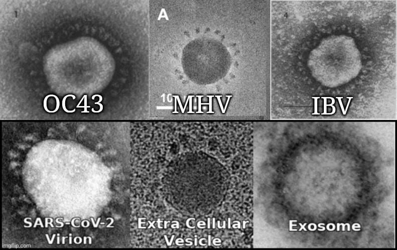
“We performed transmission electron microscopy using Abs specific for SARS-CoV-2 spike to demonstrate the presence of SARS-CoV-2 Ags on the surface of exosomes from controls and healthy vaccinated individuals. Exosomes from vaccinated individuals are positive for SARS-CoV-2 Ag (Fig. 1B). We have also stained both the exosome samples with coronavirus FIPV3-70 Ab as negative control and did not observe any positive reaction in exosomes (Fig. 1B).”

See the tiny black dots? Those are the “spikes.”
https://www.jimmunol.org/content/207/10/2405#ref-7
“Many clinically relevant viruses are simply difficult to grow or cannot be grown at all in cultured cells, while other viruses require specialized culture systems that are either not available or too complicated for routine use in diagnostic laboratories. Traditional tube cultures, although viewed as being comprehensive in growing a wide range of viruses and capable of detecting unsuspected new viruses or more common viruses in new places, fail to isolate viruses in many instances and can take days to weeks to provide a final result. While centrifugation-assisted cultures using individual, mixed, or genetically engineered cell lines are designed to be faster and more user-friendly than tube cultures, they are not always as sensitive and are normally limited by the quality and availability of reagents and the number and types of cell lines that can be used to grow a variety of different viruses.”
“Viral culture systems really have not been standardized or scrutinized to the same extent as molecular testing and can vary considerably, depending upon the selection of appropriate cell lines; the adequate collection, transport, and handling of specimens to ensure virus viability; and the maintenance of viable and healthy inoculated cells.”
https://www.ncbi.nlm.nih.gov/pmc/articles/PMC3536207/
“Examples of well-known cell types that are standard for most virology laboratories are primary rhesus monkey kidney (RhMK) cells, primary rabbit kidney cells, human lung fibroblasts (MRC-5), human foreskin fibroblasts, human epidermoid carcinoma cells (HEp-2), human lung carcinoma cells (A549), and others.”
https://www.ncbi.nlm.nih.gov/pmc/articles/PMC1797634/
“Barbara McClintock might be surprised to learn how well recent discoveries support her hypotheses. Her experiments of 60 years ago led her to propose that cells under environmental stress activate transposable elements in order to restructure the cell genome (McClintock, 1984).“ https://www.sciencedirect.com/science/article/pii/S1097276507004510
“Cell lines are used extensively in research and drug development as models of normal and cancer tissues. However, A SUBSTANTIAL PROPORTION OF CELL LINES IS MISLABELED OR REPLACED BY CELLS DERIVED FROM A DIFFERENT INDIVIDUAL, TISSUE OR SPECIES. The scientific community has failed to tackle this problem and CONSISTENTLY THOUSANDS OF MISLEADING AND POTENTIALLY ERRONEOUS PAPERS HAVE BEEN PUBLISHED USING CELL LINES THAT ARE INCORRECTLY IDENTIFIED.”
https://www.nature.com/articles/nrc2852
“WHILE PROBLEMS WITH CELL LINE MISIDENTIFICATION HAVE BEEN KNOWN FOR DECADES, AN UNKNOWN NUMBER OF PUBLISHED PAPERS REMAINS IN CIRCULATION REPORTING ON THE WRONG CELLS WITHOUT WARNING OR CORRECTION. Here we attempt to make a conservative estimate of this ‘contaminated’ literature. WE FOUND 32,755 ARTICLES REPORTING ON RESEARCH WITH MISIDENTIFIED CELLS, IN TURN CITED BY AN ESTIMATED HALF A MILLION OTHER PAPERS. The contamination of the literature IS NOT DECREASING OVER TIME and is anything but restricted to countries in the periphery of global science. The decades-old and often contentious attempts to stop misidentification of cell lines have proven to be insufficient.”
“The misidentification of cell lines is a stubborn problem in the biomedical sciences, CONTRIBUTING TO THE GROWING CONCERNS ABOUT ERRORS, FALSE CONCLUSIONS AND IRREPRODUCIBLE EXPERIMENTS [1, 2]. As a result of mislabelled samples, cross-contaminations, or inadequate protocols, some research papers report results for lung cancer cells that turn out to be liver carcinoma, OR HUMAN CELL LINES THAT TURN OUT TO BE RAT [3, 4]. In some cases, these errors may only marginally affect results; in others THEY RENDER RESULTS MEANINGLESS [4].”
“Although no exact numbers are known, THE EXTENT OF CELL LINE MISIDENTIFICATION IS ESTIMATED BETWEEN ONE FIFTH AND ONE THIRD OF ALL CELL LINES [4, 14]. (Although currently only 488 or 0.6% of over 80,000 known cell lines have been reported as misidentified, most cell lines are used infrequently [15].) In addition, MISIDENTIFIED CELL LINES KEEP BEING USED UNDER THEIR FALSE IDENTITIES LONG AFTER THEY HAVE BEEN UNMASKED [16], WHILE OTHER RESEARCHERS CONTINUE TO BUILD ON THEIR RESULTS. Considering the biomedical nature of research conducted on these cell lines, CONSEQUENCES OF FALSE FINDINGS ARE POTENTIALLY SEVERE and costly [17], with grants, patents and even drug trials based on misidentified cells [18].”
“Before any action can be taken, it is essential that we get a sense of the size and nature of the problem of contaminated literature. This raises several questions. First, HOW MANY RESEARCH ARTICLES HAVE BEEN BASED ON MISIDENTIFIED OR CONTAMINATED CELL LINES? HOW WIDE IS THEIR INFLUENCE ON THE SCIENTIFIC LITERATURE? Second, what can we say about origins and trends in the contaminated literature? Is the problem getting better, or restricted to peripheral regions of the world’s research, where perhaps protocols are less strict? Third, what could be appropriate ways to deal with the contaminated literature?”
“Using complementary search strategies (see methods), WE WERE ABLE TO IDENTIFY 32,755 ARTICLES (on August 4th, 2017) BASED ON CELL LINES THAT ARE CURRENTLY KNOWN TO BE DIFFERENT FROM THE CELL LINES REPORTED IN THESE PUBLICATIONS. As we only searched for cell lines known to be misidentified, THIS CONSTITUTES A CONSERVATIVE ESTIMATE OF THE SCALE OF CONTAMINATION IN THE PRIMARY LITERATURE.”
“In addition, RESEARCH BASED ON MISIDENTIFIED CELL LINES HAS A WIDE IMPACT ON THE SCIENTIFIC LITERATURE, AS IT APPEARS THAT THESE RESEARCH PAPERS ARE COMPARATIVELY HIGHLY CITED. WoS does not allow for precise total numbers, but we can give indications of this ‘secondary contamination’ of the literature. Analysing citations to primary contaminated articles, WE FOUND 46 PAPERS WITH MORE THAN A THOUSAND CITATIONS AND OVER 2600 CONTAMINATED ARTICLES WITH OVER A HUNDRED CITATIONS. Furthermore, OVER 92% OF THE CONTAMINATED PAPERS ARE CITED AT LEAST ONCE, which is more than average for biomedical literature [34]. In total, we can CONSERVATIVELY ESTIMATE THE CITATIONS TO THE PRIMARY CONTAMINATED PRIMARY LITERATURE AT OVER 500,000, excluding self-citations, thereby leaving traces in a substantial share of the biomedical literature. Even though it is clear that articles may receive citations for many reasons, including negative or even ritual citations, and hence not all citing articles contain (critical) errors, THE AMOUNT OF RESEARCH POTENTIALLY BUILDING ON FALSE GROUNDS REMAINS WORRISOME.”
“One might wonder whether the contamination of the research literature is mainly a problem of the past, given that the FIRST CONCERNS ABOUT MISIDENTIFIED CELL LINES WERE EXPRESSED HALF A CENTURY AGO [9, 10] and that numerous initiatives have tried to alleviate the problem since.
Based on the set of 32,755 records of primary contaminated literature, we analysed the publication dates of the articles. THE MAJORITY OF THE ARTICLES, 57%, WERE WRITTEN SINCE 2000 AND THE NUMBER OF ARTICLES USING MISIDENTIFIED CELL LINES IS STILL GROWING (see Fig 2). Clearly, the problem is definitely not one of the past, but is very relevant to contemporary science, with 58 new articles based on contaminated literature appearing even as recently as February 2017.”
“Fig 2 indicates three moments in history when cell line contamination became evident. First, through the work of Stanley Gartler it became possible to detect intraspecies cell contamination, AFTER WHICH SEVERAL OF SUCH CONTAMINATIONS INVOLVING HeLa CELLS WERE REPORTED IN NATURE IN 1968 [9, 10]. Second, cell culture contamination was put on the global research agenda by the work of Walter Nelson-Rees et al. in the 1970s [7, 8], CULMINATING IN A LIST OF CONTAMINATED CELL CULTURES IN SCIENCE IN 1981 THAT DEMONSTRATED LARGE-SCALE CONTAMINATION OF CELL CULTURES BY HeLa CELLS [44]. From this point on, it could be expected that most scientists working in those areas of research frequently employing cell cultures, were aware of the potential issues with their research material. HOWEVER, THE VAST MAJORITY OF RESEARCH PAPERS BASED ON MISIDENTIFIED CELL LINES WAS PUBLISHED AFTER THIS POINT IN TIME. Even after the introduction of STR in 2001 [45], THE ANNUAL NUMBER DOES NOT DECREASE.”
“Similar to the primary literature, the number of articles in the secondary literature is also still growing. IN 2016, OVER 40,000 PAPERS WERE PUBLISHED THAT REFERRED TO PRIMARY CONTAMINATED LITERATURE. In addition, from the information in the Supplementary Material (S2 File), we conclude that THE MAJORITY OF MISIDENTIFIED CELL LINES CONTINUE TO CONTAMINATE THE SECONDARY LITERATURE IN 2017.”
“For example, several recent publications indicate levels of CELL LINE CONTAMINATION FOR CHINA BETWEEN 25% [13] AND 46% [46] AND DEMONSTRATE THAT OF ALL ‘NEW’ CELL LINES DEVELOPED IN CHINA 85% ACTUALLY TURNED OUT TO BE HeLa CELLS [13].
However, THE MAJORITY OF THE ARTICLES USING MISIDENTIFIED CELL LINES ORIGINATE FROM COUNTRIES HOLDING WELL-ESTABLISHED RESEARCH TRADITIONS (e.g. US, Japan, Germany). Relative to their share of total research output, authors from these countries OFTEN PERFORM RESEARCH ON MISIDENTIFIED CELL LINES. In fact, mainly due to their enormous share of total literature on cell lines, OVER 36% OF ALL CONTAMINATED PRIMARY LITERATURE STEMS FROM THE US.”
“Our results seem to present worrying problems for the biomedical sciences. ALTHOUGH THE ISSUE OF MISIDENTIFIED CELL LINES HAS LONG BEEN KNOWN, ITS EFFECT ON THE SCIENTIFIC LITERATURE HAS NOT BEEN PROPERLY RECOGNIZED, LET ALONE PROPERLY TREATED [47, 48].”
“Despite measures to authenticate new and existing cell lines [27], RESEARCH BASED ON THE WRONG CELLS IS STILL PRESENT IN THE LITERATURE AND IN FACT CONTINUES TO BE PUBLISHED.”
https://journals.plos.org/plosone/article?id=10.1371/journal.pone.0186281
You won’t believe what Covid-19 PCR tests are looking for
https://rumble.com/v1f7bl1-you-wont-believe-what-covid-19-pcr-tests-are-looking-for.html


—» The Sleepy Student (Problem of stored beliefs) «—

https://www.taylorfrancis.com/books/mono/10.4324/9781003121091/epistemology-kevin-mccain

CDC deliberately lied to trick parents into vaccinating their children regardless adverse effects and inefficacy:
Side note —» Even though some parts might be similar, we should not consider the content of vaccines and the parts that make up the virus as the same. They tweaked various batches ! It’s a mess. «—
—>> “I believe it” <<— that’s why everyone is full of shit ! —> “don’t ask, just believe it”
“everyone is believing it, so should you”
“Don’t be like them”
Most people don’t favour truth over their ego and insecurities. Pathetic to claim ethical ground !
Can I suggest not to toss various people into one box?
There is a difference between "no virus" and the "classification of this virus"
Nuances do matter.
I am on about inconsistencies, no matter the topic or who it concerns.
I would never claim to know for sure what's relevant because this would mean I understand the whole picture.
Think about this:
—>> You accuse certain institutions of manipulation of data while you rely on the same institutions to make a point for another argument <<— relying on others to do the work
What is science about?

.
Thank you!!!
This is the best compilation about the decades of unbridled cell line contamination in papers before all the other research sins you pointed out are committed. I recall reading that the NIH 300 were hopelessly contaminated. I remember China blaming ALL of their fraud on the U.S. (Chinese scientists are under the gun to produce glory for the CCP. China has many journals, so it all gets published as a result). I remember a fellow with a prolific cell line business, and when the NIH 300 contamination was announced, he was contacted by customers wondering what they had paid for. Alas, he lost his entire business. He was careful, too!
I have copied and saved this with attribution to you and the url at the top and am sharing it. This is very helpful and very needed in this time of bald deceit and crimes.
Well done, Manu!